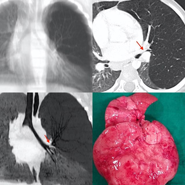
This 3 years old boy came with sudden breathlessness. A simulated radiograph from the CT scan images shows increased lucency of the left upper and mid-zones, which on the axial CT scan is seen a left upper lobe overinflation due to a high-grade stenosis of the left upper lobe bronchus (arrow). The minIP image shows this well (arrow). The operated overinflated lung is seen in the last panel.
Congenital Lobar Emphysema (CLE)
Blogs
Latest From Gallery


