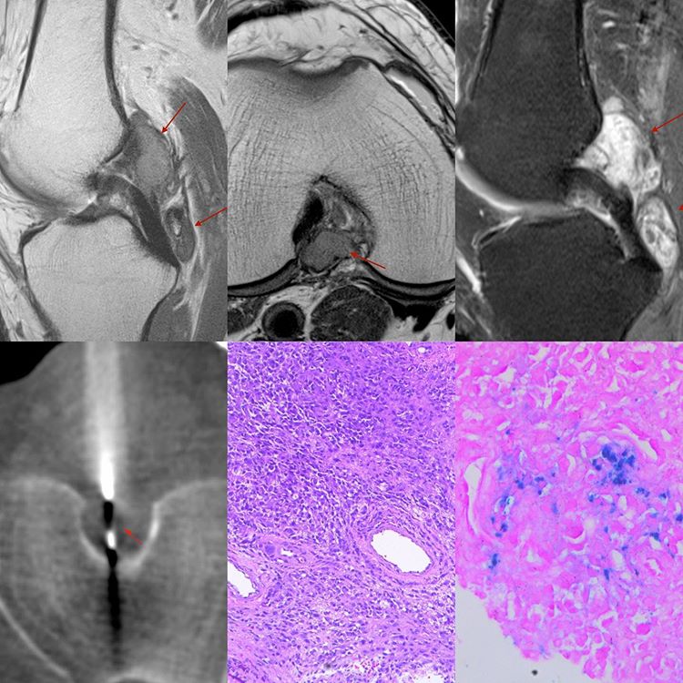
This 53-years old man presented with left knee pain.
His MRI showed a T2 intermediate and dark lesion posterior to the ACL and PCL, with homogeneous enhancement. The findings were suggestive of nodular synovitis, perhaps pigmented.
The surgeon wanted pre-treatment confirmation, so a biopsy was performed from the posterior aspect using an 18G coaxial biopsy gun.
The diagnosis was “tenosynovial giant cell tumor” (localized type), which is basically nodular synovitis (histopath images 10 x). Hemosiderin was seen on the Perls stain HP image (last image), which is the “pigmented” component.


